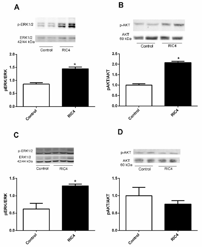
Figure 5.
Western blotting for ERK and AKT in epididymal adipose and tibial muscle tissues following ip administration of Ric4 to WT mice. Mice were treated ip with vehicle (Control) or Ric4 (600 µg/kg). Tissues were collected 30 min after the Ric4 administration and processed for Western blots as detailed in Section 2.5. The phosphorylation of either ERK or AKT in epididymal adipose tissue (A,B) and tibial muscle tissue (C,D) were analyzed using antibodies anti-pErk (A,C) or anti-pAkt (B,D). Anti-total ERK, anti-total AKT, and β-actin were used for normalizing protein concentration. Imaging and band intensity measurements were performed using ImageJ 1.49 software. Data are representative of three independent experiments that produced similar results. The statistical comparisons were performed using Student’s t-test using GraphPad Prism software * p < 0.05.

