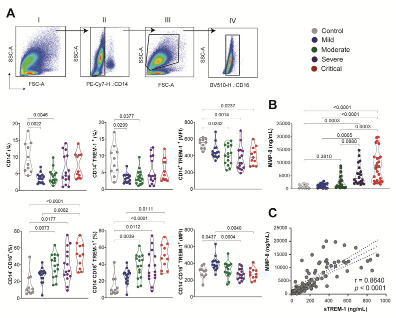
Figure 4.
sTREM-1 was released from the surface of the peripheral blood leukocyte membrane, correlated with the expressionof MMP-8. (A) Sequential gating is shown: (I) SSC-A versus FSC-A; (II) leukocytes gated according to their side scatter and CD14 antibody staining patterns; (III) light scatter flow cytometry profile for cells based on forward scatter (FSC-A) related to size, and side scatters (SSC-A) related to granularity; (IV) gated according to their side scatter and CD16 antibody staining patterns. The percentage of CD14+ and CD14− CD16+ cells was evaluated for groups of COVID-19 patients, as well as the percentage of CD14+TREM-1+ and CD14−CD16+TREM-1+ in the cell surface. The amount of TREM-1 expression was obtained by quantification of MFI in CD14+ and CD14−CD16+ leukocytes. (B) MMP-8 quantification in subgroups of COVID-19 patients. Median values are presented with ranges. The Kruskal–Wallis test was used for multiple comparisons in data with non-normal distribution. Differences between groups are indicated by the p-values in the graphics. (C) Spearman test correlation between MMP-8 and sTREM-1 levels. The correlation coefficients (r) and the p-value are indicated in the graphic.

