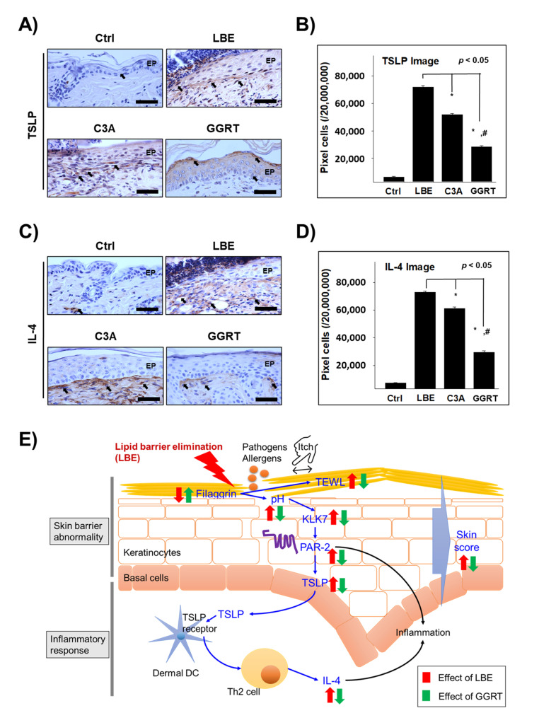
Figure 5.
The effect of GGRT on the regulation of Th2 differentiation-related proteins. Atopic dermatitis was induced by the elimination of the lipid barrier via SDS application. Isotonic sodium chloride solution, 5% ceramide, and 5% GGRT were administered to lipid-eliminated mice skins for 3 days. (A,C) TSLP and IL-4 (arrows indicate light brown particle) were visualized by immunohistochemistry using corresponding antibody. Bar size, 50 μm. (B,D) The densitometric data is shown as positively stained cells per 2 × 107 pixels of images for each protein. *, p < 0.05 compared with LBE; #, p < 0.05 compared with C3A. (E) Schematic representation of inhibition of GGRT on the LBE-induced atopic-like dermatitis. Abbreviations: Ctrl, healthy control group; LBE, normal saline-treated group after lipid barrier elimination; C3A, 5% ceramide 3B treated group after lipid barrier elimination; GGRT, 5% GGRT extract treated group after lipid barrier elimination; EP, epithelium.

