Figure 4.
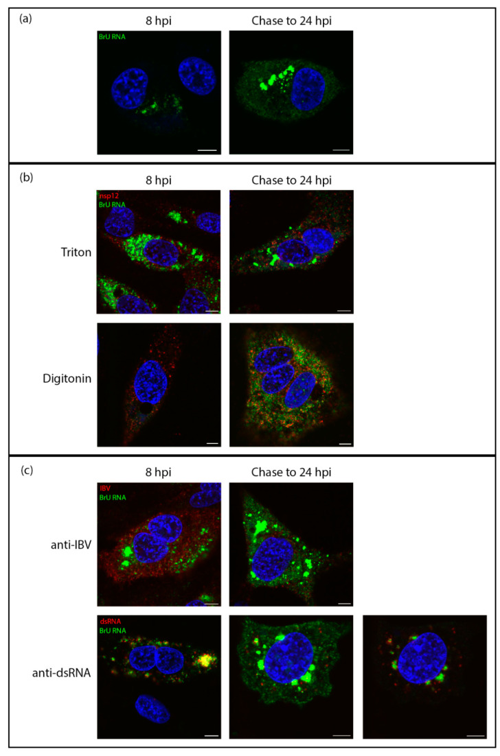
Viral RNA is transported to the cytoplasm later in infection. DF1 cells were infected with IBV. At 7 hpi cells were treated with BrU and ActD. At 8 hpi cells were either fixed (8 hpi) or chased with uridine until 24 hpi (chase to 24 hpi). (a) cells were then labeled for BrU (green), DAPI labeling the nuclei (blue). (b) DF1 cells infected with IBV were treated with BrU and ActD at 7 hpi then either fixed at 8 hpi or chased with uridine until 24hpi (chase to 24 hpi). Cells were permeabilized with Triton X-100 (top) or digitonin (bottom) then labelled for BrU (green) and nsp12 (red), DAPI labeling the nuclei (blue). (c) DF1 cells infected with IBV were treated with BrU and ActD at 7 hpi then either fixed at 8 hpi or chased with uridine until 24hpi (chase to 24 hpi). Cells were then labeled for BrU (green) and IBV (red; top) or dsRNA (red; bottom), DAPI labeling nuclei (blue). Scale bars represent 5 µm. Images are representative of three independent replicates.

