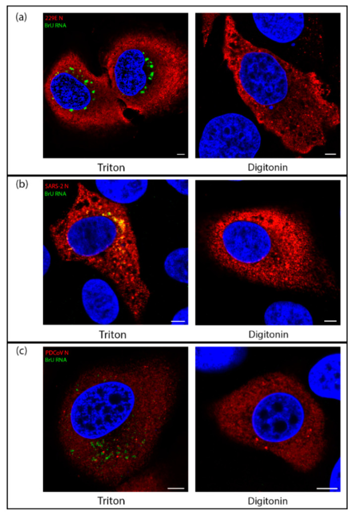
Figure 5.
Viral RNA synthesis of diverse CoVs takes place within a membrane-bound compartment. (a) Huh7 cells were infected with HCoV 229E. At 7.5 hpi cells were treated with BrU and ActD for 30 min before fixation at 8 hpi. Cells were permeabilized with Triton X-100 (left) or digitonin (right) then labeled for BrU (green) and N (red), with DAPI labeling the nuclei (blue); (b) VeroE6 cells were infected with SARS-CoV-2. At 5.5 hpi, cells were treated with BrU and ActD for 30 min before fixation at 6 hpi. Cells were permeabilized with Triton X-100 (left) or digitonin (right) then labeled for BrU (green) and N (red), with DAPI labeling the nuclei (blue); (c) LLC-PK1 cells were infected with PDCoV. At 5.5 hpi, cells were treated with BrU and ActD for 30 min before fixation at 6 hpi. Cells were permeabilized with Triton X-100 (left) or digitonin (right) then labeled for BrU (green) and N (red), with DAPI labeling the nuclei (blue). Scale bars represent 5 µm. Images are representative of three independent replicates.

