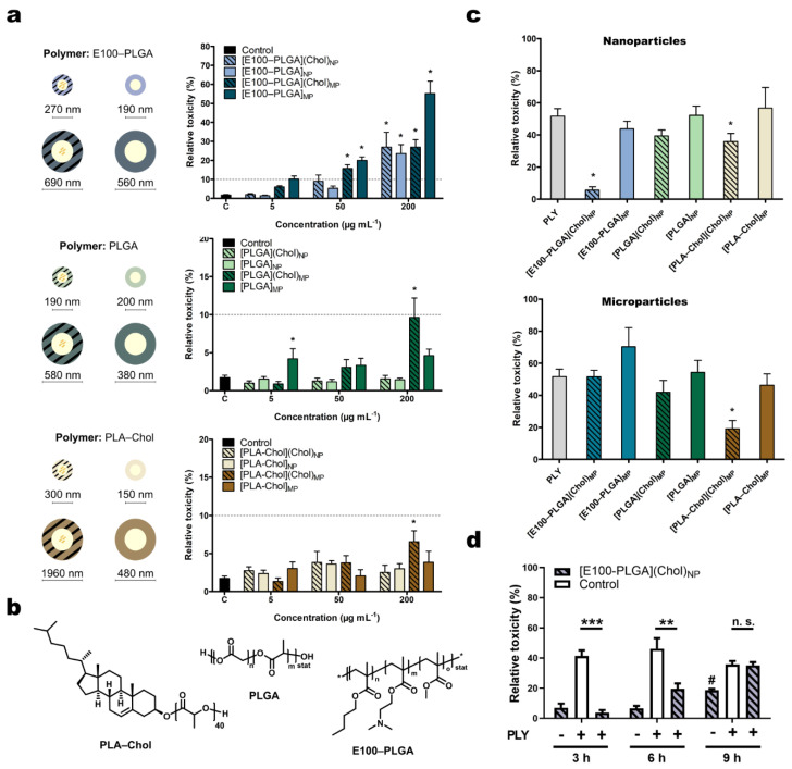
Figure 1.
Evaluation of cell toxicity after polymer particle stimulation or pneumolysin stress. (a) HepG2 cells were treated with polymeric particles prepared by nano- or microprecipitation (NP or MP, respectively) of indicated concentrations in DMEM:F12 for 3 h. Stripes indicate the cholesterol cargo of the polymer particle. Mean diameter size (rounded) of tested sample and polymer composition (b) after lyophilization is shown. The polymer surface was coated with poly(2-oxazoline) (POx). (c) Cells were challenged with PLY (250 ng mL−1) for 3 h in the presence of nano- or microprecipitated particles (50 µg mL−1) before toxicity was examined by the release of cytoplasmic LDH. (d) Amelioration of PLY toxicity by [E100–PLGA](Chol)NP was found to be time-dependent. Mean ± SEM toxicity is shown relative to completely lysed cells (= 100%). * p < 0.05, ** p < 0.01, *** p > 0.001, one-way ANOVA, corrected for multiple comparisons against untreated control (a) or PLY (c,d) (Dunnett’s test).

