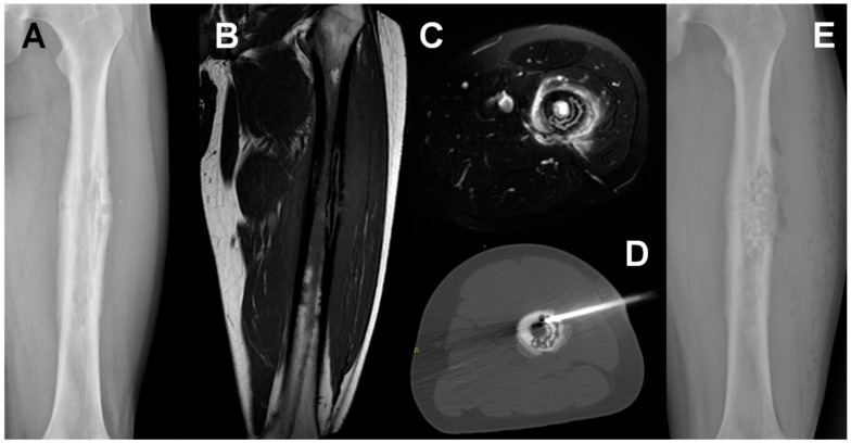
Figure 1.
Staphylococcus aureus Osteomyelitis in a 20-year-old man. A conventional radiograph (A). MRI coronal T1w (B) and axial T2w fat-saturated (C) show a permeative lesion of the left femoral shaft. CT-guided biopsy permitted to identify the responsible microorganism (D). Conventional radiograph after surgical treatment showed antibiotic microspheres placed into the bone (E).

