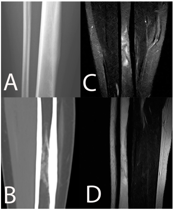
Figure 3.
Chronic osteomyelitis of the tibia in a 16-year-old female. Periosteal reaction and sclerotic intramedullary focus are detectable on conventional radiography (A) and CT scan (B). MRI showed ill-defined bone edema among the sclerotic intramedullary changes on STIR coronal (C) and T1w sagittal (D).

