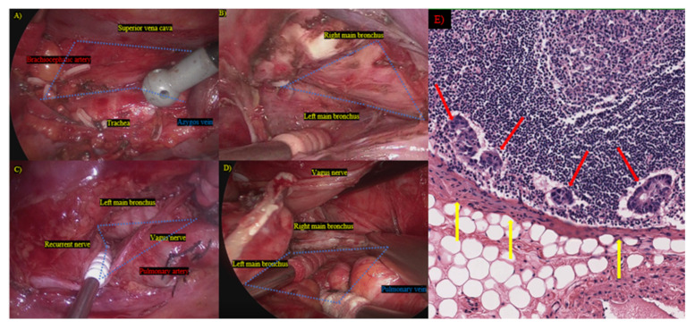
Figure 2.
Mediastinal lymph node dissection and pathological findings. (A) Right upper mediastinum; (B) right bifurcation; (C) left upper mediastinum; (D) left bifurcation; blue area: mediastinal dissected region; (E) cancer cells disseminate from metastasized lymph nodes through the sheath of the lymph nodes; red arrow: metastasized lymph node; yellow arrow: disseminated cancer cells.

