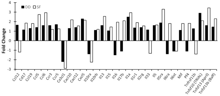
Figure 6.
Cytokine gene activation in the mPFC by SF and DD. Mice were subjected to 10 days of disturbance either during the light or dark period. Mice were then euthanized and mPFC chemokine and proinflammatory cytokine profiles determined. Brain tissue for each group was pooled (4–5 mice/group), RNA isolated and cytokine profiles determined by qPCR array. Values represent changes in RNA levels relative to undisturbed mice. SF: sleep fragmented; DD: dark disturbed.

