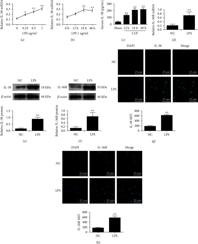
Figure 1.

Expression of IL-38 and its receptor Il-36R in mouse macrophages treated with LPS. (a) IL-38 normalized mRNA expression in mouse macrophages stimulated with LPS at the indicated concentrations for 12, 24, and 48 h. β-Actin served as the housekeeping gene. (b) IL-38 normalized mRNA expression in mouse macrophages stimulated with 1 μg/ml LPS for 12, 24, or 48 h. β-Actin was used as the housekeeping gene. n = 3; ∗P < 0.05 and∗∗P < 0.01 vs. the normal control (NC) group. NC was PBS treatment. (c) Serum concentrations of IL-38 in mice at the indicated timepoints after cecal ligation and puncture (CLP), based on ELISAs. n = 6; ∗∗P < 0.01vs. the sham group; (d) IL-36R normalized mRNA expression in mouse macrophages stimulated with LPS (1 μg/ml; 24 h). β-Actin was used as housekeeping gene. (e, f) Representative western blot image (left panels) and quantification (right panels) of IL-38 and IL-36R protein levels of macrophages stimulated with 1 μg/mL LPS for 24 h. β-Actin protein levels were used for normalization. (g, h) Representative immunofluorescence images of IL-38 (green) and IL-36R (green) of macrophages treated as in panels (d, e). Nuclei were stained with DAPI (blue). Magnification, ×600. Scale bar, 25 μm. ∗∗P < 0.01vs. the NC group. Results are shown for three independent experiments.
