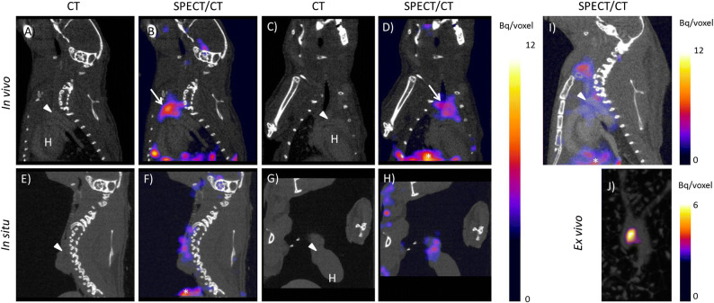
Fig. 1.
[111In]In-DOTA-JR11 uptake in mouse atherosclerotic plaque two hours post injection of 200 pmol [111In]In-DOTA-JR11. A Sagittal CT, B saggital SPECT/CT, C coronal CT, and D coronal SPECT/CT image of [111In]In-DOTA-JR11 uptake in vivo in an atherosclerotic mouse. E Sagittal CT, F saggital SPECT/CT, G coronal CT, and H coronal SPECT/CT image of [111In]In-DOTA-JR11 uptake in situ in the mouse displayed in (A-C) scanned post-mortem after thymectomy and flushing of the vasculature with PBS. I Sagittal SPECT/CT image of a mouse two hours post injection of 50 MBq/200 pmol [111In]In-DOTA-JR11 plus a 100 × excess of unlabeled DOTA-JR11. Plaque uptake was strongly reduced by blocking. J Maximum intensity projection image of the excised arteries of the mouse shown in (A-H). Focal uptake of [111In]In-DOTA-JR11 is visible at the plaque location. Arrowheads indicates the location of the aortic arch containing plaque, arrows indicates thymic uptake of [111In]In-DOTA-JR11, *Indicates uptake in the liver, H indicates the heart

