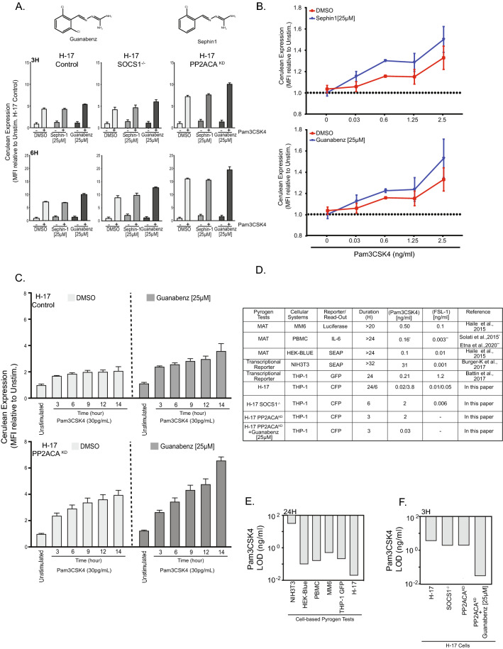
Figure 6.
PP1 inhibition potentiates the response of H-17 PP2ACAKD towards lipoprotein. (A) H17 Control, H-17 SOCS1−/−, and H-17 PP2ACAKD_25 cells were stimulated with 100 ng/ml Pam3CSK4 in the presence of DMSO (solvent control), Sephin-1 (25 µM) or Guanabenz (25 µM). Cerulean expression was determined by flow cytometry 3 h (upper panel) or 6 h (Lower panel) after stimulation. Bars represent MFI ± SEM from three independent experiments. (B) Cells were treated as in (A) and stimulated for 6 h with the indicated concentrations of Pam3CSK4. Data points represent represent MFI ± SEM (n = 4). (C) H17 Mock or H-17 PP2ACAKD were treated with DMSO or Guanabenz (25 µM) and stimulated with Pam3CSK4 (2 ng/ml) for 6, 9, 12 and 14 h respectively. Bar graphs represent MFI ± SEM (n = 4). (D) Comparison of different human cell-based pyrogen tests with regard to the limit of detection (LOD) for the TLR1/TLR2 ligand Pam3CSK4 or the TLR2/TLR6 ligand FSL-1. The respective read-out and the duration of stimulation is indicated. (E) LOD for Pam3CSK4 by different reporter cell lines upon 24 h of stimulation. LOD values were taken from references cited in (D). (F) Limit of detection (LOD) for Pam3CSK4 by H-17 reporter cells and derived knock-out and knock-down cell lines upon 3 h of stimulation.

