Figure 1.
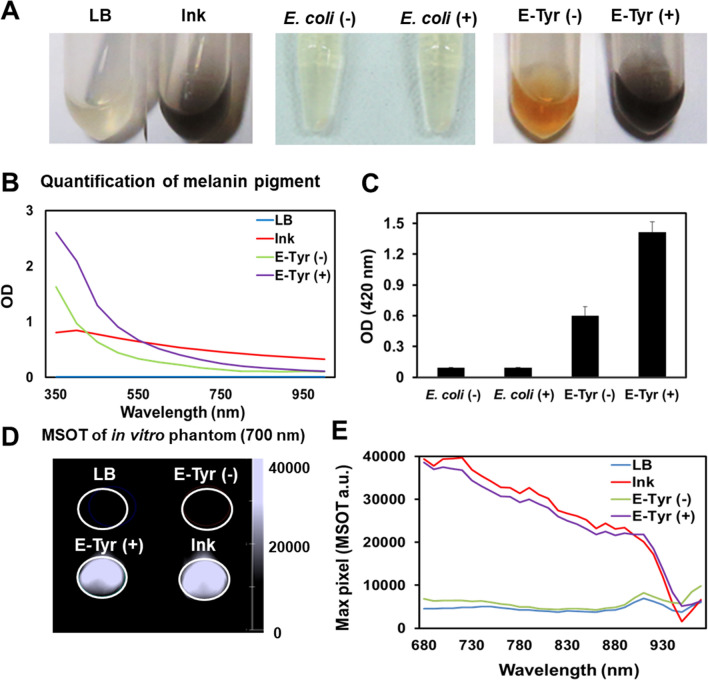
Production of melanin by E-Tyr in vitro. (A) Melanin production in vitro. Tyrosinase-expressing E. coli (E-Tyr) were grown overnight in the presence or absence of L-tyrosine supplementation. (left) LB (growth medium) and ink control, (middle) supernatant of E. coli with the tyrosinase-expression vector (control bacteria) cultured with (E. coli(+)) or without (E. coli(−)) L-tyrosine supplementation, and (right) supernatant of E. coli carrying the tyrosinase-expression vector cultured with (E-Tyr(+)) or without (E-Tyr(−)) l-tyrosine supplementation. (B) Optical density measurements of LB, ink, E-Tyr (−) and E-Tyr (+) over the spectrum from 350 to 1000 nm. (C) Quantification of melanin pigment shown in (A), E. coli (−), E. coli (+), E-Tyr (−) and E-Tyr (+), using a spectrophotometer at 420 nm wavelength. (D) Optoacoustic in vitro phantom imaging of (C) with an optoacoustic image spectrum peaked at 700 nm. (E) Optoacoustic spectra for E-Tyr with or without L-tyrosine supplementation, ink and LB media, measured between 680 and 970 nm.

