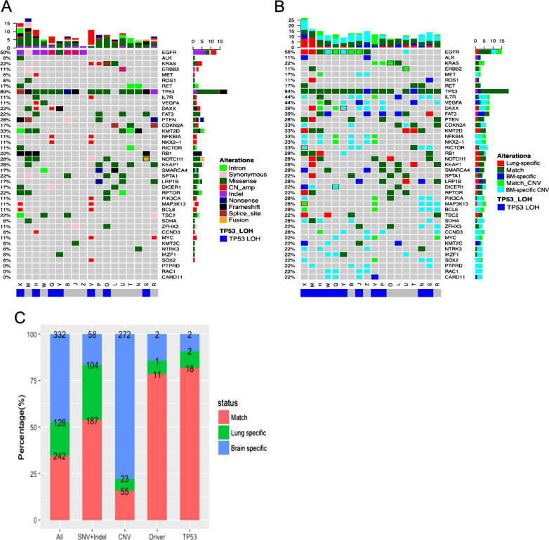
Fig. 2.
Somatic mutations in paired primary lung and brain metastatic lesions. A Genomic profiles of primary lung lesions (n = 18). Different colors denote different mutation types. B Somatic mutation profiles of paired brain metastatic lesions compared with those of primary lung lesions. Different colors denote whether mutations detected in brain metastatic tissue match with those in the paired primary lung tumor tissue (match), or were lung tumor tissue-specific or brain metastasis tissue-specific. TP53 loss of heterozygosity (LOH) status is indicated at the bottom of the Oncoprint. The top bars summarize the number of mutations carried by a patient. The left side of the oncoprint indicates the percentage of patients with mutations in the genes indicated on the right side. The side bars summarize the total number of mutations detected and the distribution of mutation types per gene. C Percentages and numbers of mutations in paired primary lung and brain metastatic lesions for the indicated mutation types. BM brain metastatic, TP53 tumor protein p53, AF allele frequency, Lung-specific, mutations only detected in primary lung lesion, match positive in the DNA of both primary and brain metastatic lesions, BM-specific mutations only detected in brain metastatic lesion, CNVs copy number variations, SNVs single-nucleotide variations, indel insertion-deletion variations

