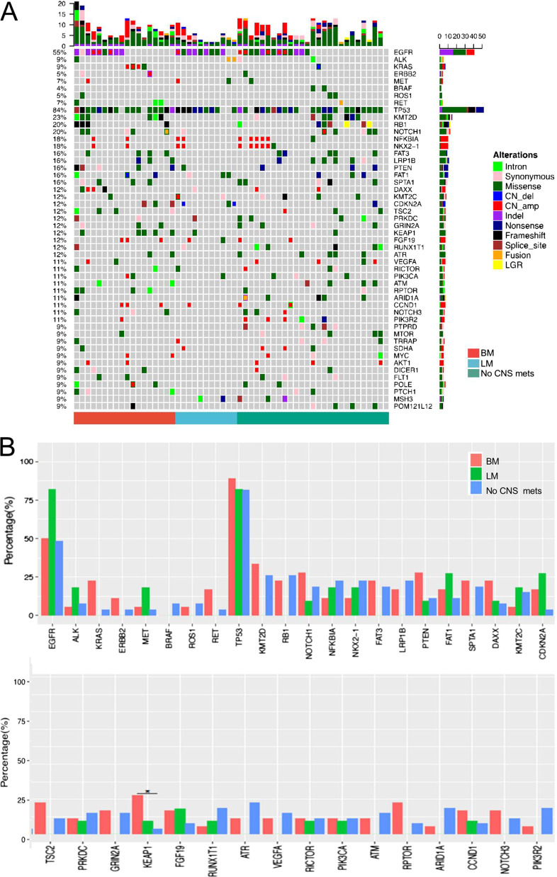
Fig. 5.
Somatic mutations of primary lesions cannot differentiate patients with and without CNS metastases. A Genetic profiles of primary lung lesions of patients with brain metastasis (n = 18), leptomeningeal metastasis (n = 11) or without CNS metastases (n = 27) as annotated below. Tissue DNA from four patients without CNS metastasis failed quality control and were excluded from this analysis. Different colors denote different mutation types. The top bars summarize the number of mutations detected from each patient. The left side of the oncoprint indicates the percentage of patients with mutations in the genes indicated on the right side. The side bars summarize the total number of mutations detected and the distribution of mutation types per gene. B Rate of detection for the indicated genes. *P < 0.05. CNS central nervous system, BM brain metastasis, LM leptomeningeal metastasis, no CNS metastases, patients who never developed CNS metastases before their cancer-related death; *P = 0.031 KEAP1 detected in patients with BM vs. those with no CNS metastases. Analyzed by Fisher’s exact test

