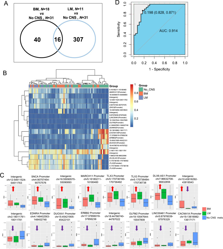
Fig. 6.
Differentially methylated blocks in patients with and without CNS metastasis. A–C 15 methylation blocks identified to be differentially methylated in primary lung tumor samples from patients with either brain or leptomeningeal metastasis as compared to patients without CNS metastasis. A Venn diagram showing the 16 differentially methylated blocks shared between patients with either brain or leptomeningeal metastases. B Heat map summarizing the unsupervised hierarchical clustering of methylation profiles of each patient for the 16 methylation markers consistently identified to be differentially methylated in primary lung lesions of patients with either brain or leptomeningeal metastasis. Differentially methylated blocks were identified using unpaired two-tailed t-tests with P < 0.05. C Box plots plotting the methylation values (y-axis) and showing the hypermethylation (red arrow pointing upwards) or hypomethylation (purple arrow pointing downwards) patterns for the 16 DNA methylation markers of the cohort. Fifteen blocks have identical patterns in patients with either brain or leptomeningeal metastases; 1 block is hypermethylated in brain metastasis but hypomethylated in leptomeningeal metastases. D Receiver Operating Characteristic curves were generated to assess the performance of the predictive model using the 15 methylation markers to distinguish between patients with and without CNS metastasis. CNS central nervous system, BM brain metastases, LM leptomeningeal metastases, no CNS metastasis, patients who never developed CNS metastasis before their cancer-related death, AUC area under curve

