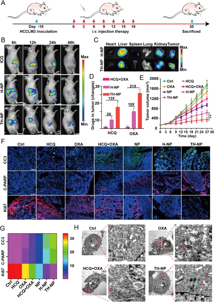
Fig. 4.
TH-NPs enhanced chemotherapy on HCCLM3 subcutaneous tumor models via autophagy inhibition. A Schedule of the antitumor treatment on subcutaneous HCCLM3 xenograft models. Tumor bearing mice (n = 8) with the volume about 150 mm3 received i.v. injection of the indicated drugs every three days for seven times. The injection dosages at equivalent dosage of OXA (10 mg/kg) or HCQ (20 mg/kg) in free drugs or NPs, H-NPs or TH-NPs. Mice treated with saline was served as control. Tumor volumes were measured for 27 days and sacrificed for the next analysis on day 30. B In vivo IVIS imaging was performed to monitor the biodistribution of TH-NPs on subcutaneous tumor-bearing nude mice (n = 3). Mice were intravenously injected with free ICG (1.5 mg/kg), or equivalent ICG-labeled H-NPs or TH-NPs and the FL images were obtained at 6, 12, 24 and 48 h post injection. C Ex vivo IVIS imaging was used to examine the ICG FL signals of major organs and tumor to further study the biodistribution of the nanoparticles, after the mice were sacrificed at 48 h post injection. D OXA and HCQ content in the tumor tissues. On day 18, three mice in OXA/HCQ combination, H-NPs or TH-NPs group were sacrificed at 24 h post i.v. injection to compare the content of OXA and HCQ. E Growth curves of tumors after different treatments. TH-NPs had the most significant tumor growth limitation compared to other groups. F Immunohistochemical frozen sections (IHC-F) of tumor tissues of the different groups and G its heat map of quantitative analysis. Scale bar: 200 μm. H TEM images of the autophagic vacuoles in the tumor tissues of different groups. (N, nucleus; red arrow, autolysosomes; green arrow, autophagosomes). Data were displayed as the mean ± SD. *P < 0.05; **P < 0.01; ***P < 0.001

