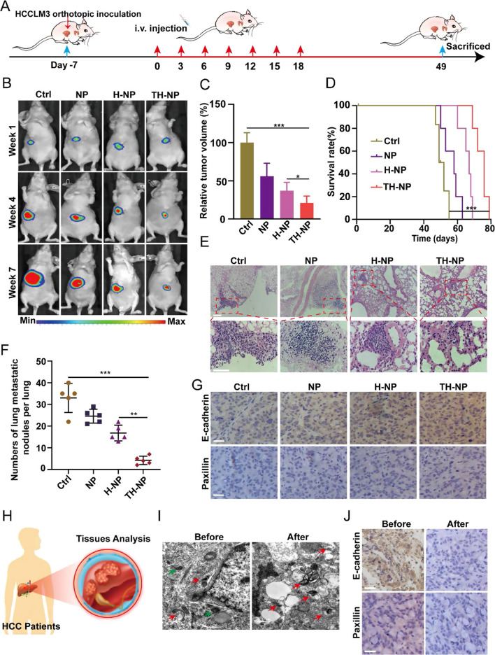
Fig. 5.
TH-NPs inhibited tumor metastasis by reversing FAs disassembly in orthotopic HCC models. A Schedule of the treatments on HCCLM3 orthotopic tumor models. B Orthotopic HCCLM3 tumor sites (n = 4) at the indicated week were localized by bioluminescence-based imaging. Mice received i.v. injection of saline, NPs, H-NPs, or TH-NPs at equivalent dosages (OXA: 10 mg/kg; HCQ: 20 mg/kg). C The relative tumor volume was quantified by bioluminescence intensity, and TH-NPs treatment showed the highest orthotopic tumor inhibition. D Kaplan–Meier survival plot of HCCLM3 orthotopic tumor-bearing mice after i.v. injection of the indicated formulations. E H&E staining of lung metastatic nodules of HCCLM3 tumor-bearing mice at the end of the experiments, Scale bar: 100 μm. F The numbers of lung metastatic nodules in the lungs. G IHC staining showing focal adhesions including E-cadherin and Paxillin increase after TH-NPs treatments, which limited tumor distant metastasis. Scale bar: 100 μm. H Scheme showing the cancer tissues of HCC patients were obtained based on the following analysis. I TEM images of the autophagic vacuoles in the HCC patients before or after chemotherapy. (N, nucleus; red arrow, autolysosomes; green arrow, autophagosomes). Scale bar: 0.5 μm. J IHC staining of E-cadherin and paxillin. The patients who received OXA-based chemotherapy (after) had a low expression compared to the patients without chemotherapy (before). Scale bar: 100 μm. Data are displayed as the mean ± SD. *P < 0.05; **P < 0.01; ***P < 0.001

