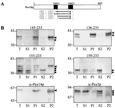
FIG. 8.
The central matrix loop of Pex13p interacts tightly with the peroxisome membrane. (A) Schematic presentation of the Pex13p-deletion mutants fused to the N terminus of GFP (*). The TMDs are shaded in black, and residue numbers are on the left. (B) CHO cells were transiently transfected with plasmids expressing one of the deletion proteins schematically presented in panel A. After 24 h, the cells were fractionated as described in Materials and Methods. Equivalent portions of the total (T), the buffer A-soluble (S1), the buffer A-insoluble (P1), the carbonate-soluble (S2), and the carbonate-insoluble (P2) material were separated by sodium dodecyl sulfate-polyacrylamide gel electrophoresis and immunoblotted with an antiserum raised against GFP. Similar fractions obtained from nontransfected CHO cells were probed with anti-Pex14p antiserum, an antiserum that specifically recognizes the integral PMP Pex14p (11), and anti-Pex5p antiserum, an antiserum that specifically recognizes the predominantly cytosolic PTS1-protein import receptor Pex5p. Arrows, the GFP fusion proteins Pex5p and Pex14p; ●, degradation products. The migrations of the molecular mass markers (masses shown in kilodaltons) are indicated.

