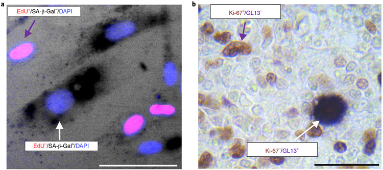
Fig. 3 ∣. Double staining SA-β-Gal or LF and proliferation markers.
a, Human BJ fibroblasts were treated with 4 μM vorinostat (SAHA) for 8 d. Drug was refreshed daily. Eight days post-treatment, mutually exclusive staining between SA-β-Gal and EdU was observed. The purple arrow denotes an EdU+/SA-β-Gal− cell and the white arrow depicts an EdU−/SA-β-Gal+ (senescent) cell. Scale bar, 150 μm. b, Double immunohistochemical/hybrid histo-immunohistochemical staining in human primary classical Hodgkin lymphomas (cHLS). A mutually exclusive staining pattern between GL13 and Ki-67 in neoplastic Hodgkin and Reed-Sternberg cells was observed. The purple arrow denotes a Ki-67+/GL13− cell and the white arrow depicts a Ki-67−/GL13+ (senescent) cell. Alkaline phosphatase (AP) chromogenic reaction: dark blue cytoplasmic product; diaminobenzidine (DAB) reaction: nuclear brown signal; counterstain: nuclear fast red. Scale bar, 50 μm.

