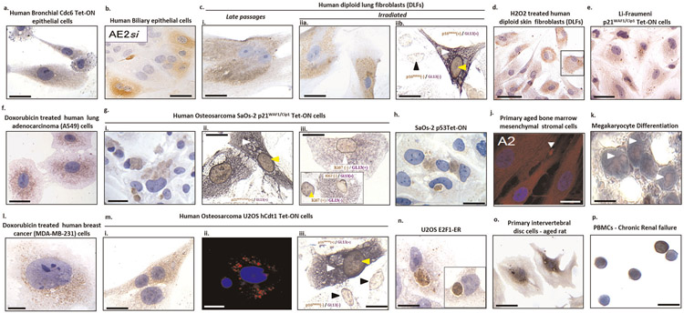
Extended Data Fig. 1∣. Sensitive detection of senescence in a variety of cell types using GL13 in cellular systems.
Positive GL13 staining is depicted in normal (a and b) and cancerous (f and l) cells of epithelial origin, normal (c, d, j, o), premalignant (e) and malignant mesenchymal (g, h, m, n) cells as well as in differentiating megakaryocytes (k)61 and peripheral blood mononuclear cells (PBMCs) from patients suffering from a chronic aged-related disease (p). Chromogenic assays: Diaminobenzidine (DAB)-brown cytoplasmic signal (a, b, ci-ii, a, d-gi, h, l, mi, n-p) and Alkaline phosphatase (BCIP/NBT)-blue purple cytoplasmic signal (white arrowheads in ciib, gii, k); Fluorescent assay: granular red cytoplasmic signal (j, mii -white arrowheads). Double staining experiments showing nuclear p16INK4A or p21WAF1/Cip1 expression (yellow arrowheads) in senescent cells that are concurrently positive with GL13 (white arrowheads) (ciib, gii and miii). Black arrowheads depict double negative cells. giii: Mutually exclusive staining pattern between nuclear Ki67 positivity (yellow arrowhead) and GL13 staining (white arrowhead). Images adopted from: a, c, g, h, m and n28; b62; j63. Counterstain: Hematoxylin (chromogenic assay) and DAPI (fluorescent assay). Scale bars: 5 μm (l), 10μm (a-c, f-k-m-p,), 15μm (e) and 20 μm (d).

