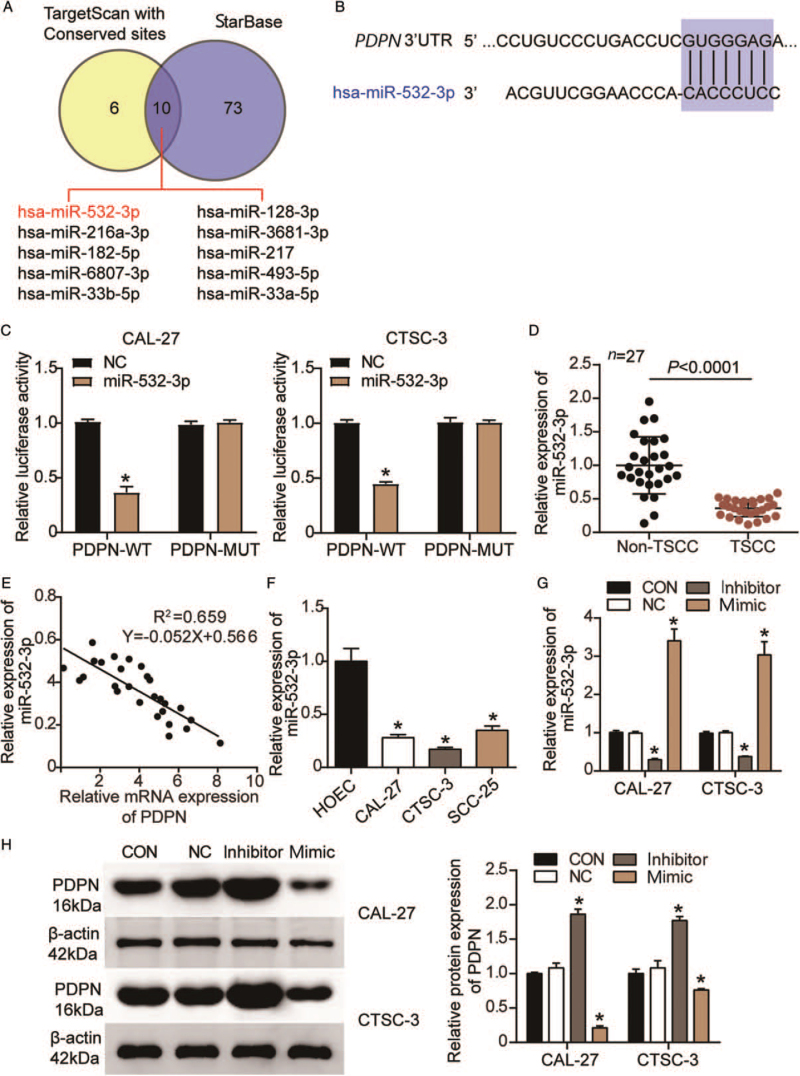
Figure 4.
PDPN was identified as a target gene of miR-532-3p in TSCC cells. (A): Ten miRNAs targeting PDPN overlapped between TargetScan and starBase. (B): The predicted binding sites for miR-532-3p on the PDPN 3′UTR. (C): A DLR assay was performed to verify the binding of miR-532-3p to PDPN. WT: Wild-type; MUT: Mutant; NC: miR-532-3p mimic NC; ∗P < 0.001 vs. the miR-532-3p mimic NC. (D): The relative expression of miR-532-3p in TSCC tissues and corresponding non-TSCC tissues as determined by RT-qPCR. (E): The correlation between miR-532-3p and PDPN in TSCC tissues was analyzed by Pearson's correlation. (F): The relative expression of miR-532-3p in the HOEC and TSCC cell lines (CAL-27, CTSC-3, and SCC-25) as determined by RT-qPCR. (G): The transfection efficiency of the miR-532-3p inhibitor and mimic in CAL-27 and CTSC-3 cells. ∗P < 0.001 vs. the blank control group. (H): The protein expression of PDPN in CAL-27 and CTSC-3 cells after the transfection of the miR-532-3p inhibitor and mimic. CON: Blank control group; DLR: Dual-luciferase reporter; HOEC: Human normal oral epithelial cell; miRNAs: MicroRNAs; NC: Negative control; PDPN: Podoplanin; RT-qPCR: Quantitative real-time reverse transcription-polymerase chain reaction; TSCC: Tongue squamous cell carcinoma. ∗P < 0.001 vs. the blank control group.

