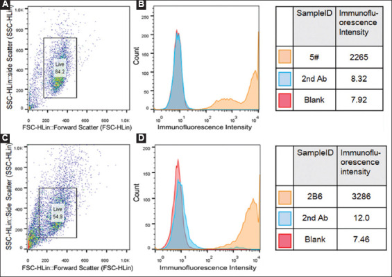
Figure 1. Construction of CHO-K1 Siglec-15 cell line. (A and B) Primary screening of CHO-K1 Siglec-15 cell lines by flow cytometry. (C and D) Secondary screening of CHO-K1 Siglec-15 cell lines by flow cytometry. The primary antibody was 5G12 and the secondary antibody (2nd Ab) was PE Goat anti-human IgG Fc. Cells were stained by PE. The 2nd Ab was used as the negative control. In Figure 1B and 1D, the cells expressing Siglec-15 were counted (Y axis), and the yellow fluorescence intensity (X axis) was positively related to the Siglec-15 expression.

