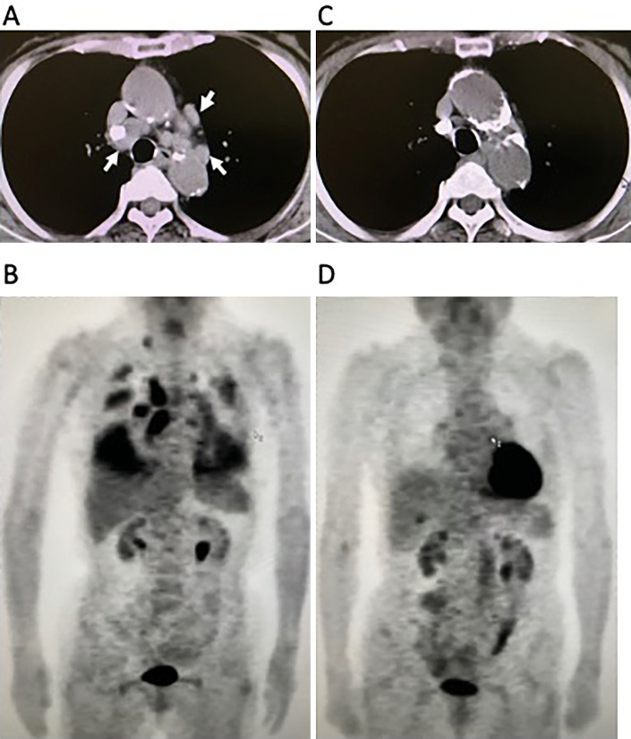
Figure 1.
Chest CT and whole-body FDG-PET findings. (A, B) Initial CT and FDG-PET findings showed abnormal hilar masses in the mediastinum (arrowheads) (A), and an abnormal uptake in the mediastinum and lung, which partially suggest carcinomatous pleuritis, and right subclavicular lesions (B). (C, D) Seven years after the first examination, chest CT and FDG-PET revealed the absence of the original abnormal hilar lesion (C), and no evidence of abnormal uptake (D). CT: computed tomography, FDG-PET: 18F-fluorodeoxyglucose-positron emission tomography

