FIG. 1.
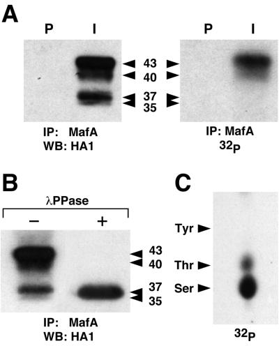
MafA is phosphorylated in HeLa cells. (A) HeLa cells transfected with pcDNA3-MafA were metabolically labeled with [32P]orthophosphate, and cellular extracts were immunoprecipitated with either preimmune (P) or MafA-specific (I) serum. Immunoprecipitates were analyzed by Western blotting followed either by incubation with anti-HA1 antibody (left panel) or by direct autoradiography (right panel). (B)MafA immunoprecipitates from HeLa cells were treated with λ phosphatase (+) or left untreated (−) and analyzed by Western blotting with anti-HA1 antibody. Apparent molecular masses (kDa) of MafA bands are indicated. (C) Phosphoamino acid analysis of 32P-labeled MafA immunoprecipitated from HeLa cells. The migration of phosphoamino acid standards is indicated.

