Figure 1.
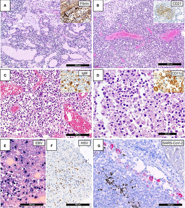
Lymph Node Histomorphology in COVID-19. (A) Overview of a lymph node draining a COVID-19 lung with edema and capillary stasis (H&E; 100x); inset: fibrin microthrombus in a dilated subcapsular sinus (immunoperoxidase; 100x). (B) Severe capillary stasis and expansion of the paracortex without discernable germinal centers (H&E; 100x); inset: disrupted, CD21+ germinal center network (immunoperoxidase; 100x). (C) Proliferation of extrafollicular plasmablasts (H&E; 200x); inset: expression of IgM by plasmablasts (immunoperoxidase; 360x). (D) Hemophagocytosis in the sinus of a lymph node (H&E; 360x); inset: positivity for CD11c in histiocytes with hemophagocytosis (immunoperoxidase; 400x). (E) Increased amount of EBV-infected B-cells in a lymph node of a COVID-19 patient (EBER ISH; 280x). (F) Increased amount of HSV infected B-cells (immunoperoxidase; 280x). (G) SARS-CoV-2 positivity in sinus histiocytes as detected by immunohistochemistry for the SARS-CoV-2 N-antigen (immunoperoxidase with 3-amino-9-ethylcarbazole used as a chromogen; 280x).

