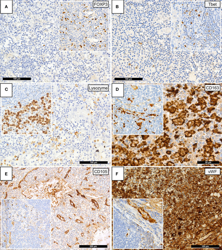
Figure 2.
Immunohistochemical Patterns of a Dysregulated Immune Response in COVID-19. Insets display immunohistochemical stainings in controls. (A) Paucity of FOXP3-positive Tregs in COVID-19 patients compared to controls (immunoperoxidase; 280x). (B) Reduced amounts of Tbet positive T-helper 1 (TH1) cells in COVID-19 versus controls (immunoperoxidase; 280x). (C) Reduced amounts of lysozyme-positive histiocytes/macrophages in the paracortex of COVID-19 lymph nodes versus controls (immunoperoxidase; 280x). (D) Increased amounts of CD163-positive histiocytes/macrophages in the paracortex of COVID-19 lymph nodes versus controls (immunoperoxidase; 400x). (E) Increased CD105-positive microvessel density in COVID-19 lymph nodes versus controls (immunoperoxidase; 50x). (F) Increased amount of vWF expression in COVID-19 lymph nodes, vWF positivity in controls is limited to the vascular wall (inset) (immunoperoxidase; 280x).

