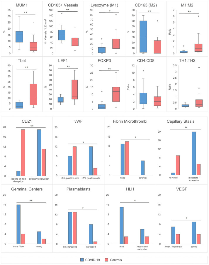
Figure 3.
Quantitative Histomorphological and Immunohistochemical Characteristics between COVID-19 Cases and Controls. A significantly higher % of MUM1+ plasmablasts, CD105+ capillaries and CD163+ macrophages is observed in COVID-19. In contrast, the % of lysozyme positive macrophages, Tbet, LEF1 and FOXP3+ T-cells was decreased. The M1:M2 (lysozyme:CD163) ratio is decreased, while CD4:CD8 and TH1:TH2 (Tbet : GATA3) ratios remain unchanged. An increased incidence of CD21+ FDC network disruption with an accompanied lack of germinal centers, increase of plasmablasts, microthrombosis, stasis/edema and moderate/extensive hemophagocytosis is observed in COVID-19 versus controls. The incidence of >5% vWF positive cells and the expression of VEGF is increased. *significant at the 0.05 level, ** significant at the 0.01 level. HLH designates increased hemophagocytic activity of histiocytes, but not hemophagocytic lymphohistiocytosis.

