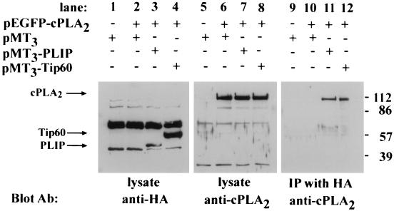
FIG. 3.
PLIP interacts with cPLA2. COS cells were transfected as follows: empty pMT3 vector alone; pEGFP-cPLA2 and pMT3; pEGFP-cPLA2 and pMT3-PLIP; and pEGFP-cPLA2 and pMT3-Tip60. Cell lysate proteins were separated by SDS-polyacrylamide gel electrophoresis (PAGE) and analyzed by Western blot analysis. Western blot with anti-cPLA2 antibody demonstrates a band at approximately 112 kDa in lanes 6, 7, and 8, which contain lysates of pEGFP-cPLA2-transfected COS cells (arrow). Western blot with anti-HA antibody demonstrates a band at approximately 50 kDa in lane 3, which contains the lysate of pMT3-PLIP-transfected cells (arrow), and at 60 kDa in lane 4, which contains the lysate of pMT3-Tip60-transfected COS cells (arrow). After immunoprecipitation of lysates with anti-HA antibody and resolution of the precipitants by SDS-PAGE, Western blot analysis with the anti-cPLA2 antibody revealed a band corresponding to GFP-cPLA2 in lysates of cells in which GFP-cPLA2 was coexpressed with HA-PLIP (lane 11) and HA-Tip60 (lane 12) but not in lysates of cells transfected with pEGFP-cPLA2 and pMT3 (lane 10).

