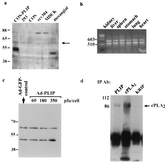
FIG. 5.
Detection and coimmunoprecipitation of Tip60-PLIP and cPLA2 in cultured mesangial cells. (a) Western blot of lysates from COS, 293, ecv304, MDCK, and mesangial cells using a polyclonal antibody raised in rabbits against GST-PLIP. A band at approximately 50 kDa is present in the lane containing lysates of HA-PLIP-transfected COS cells. A band of approximately similar size is present in the lane corresponding to rat renal mesangial cells but not in those lanes containing protein from 293, ECV304, MDCK, or nontransfected COS cells. There is a light band in each lane at 60 kDa which likely reflects Tip60. (b) Gel electrophoresis of DNA obtained by RT-PCR of RNA harvested from BALB/c mouse tissue. Two bands of 456 and 300 bp, representing Tip60 and PLIP DNA, respectively, were obtained using primers which flanked exon 5. Molecular mass markers are indicated. (c) Western blot of rat neonatal myocytes infected with Ad-GFP or with increasing amounts of Ad-PLIP. Two bands are present in all lysates at approximately 60 and 50 kDa, representing Tip60 and PLIP, respectively. Signal corresponding to PLIP is enhanced in lysates of Ad-PLIP-infected myocytes at 350 PFU/cell. (d) Mesangial cell lysate was immunoprecipitated over 4 h with anti-PLIP antibody versus a control antibody which recognizes the nuclear protein, KRIP (43), or with anti-cPLA2 antibody. Immunoprecipitants separated by SDS-polyacrylamide gel electrophoresis and transferred to an Immobilon membrane were blotted with cPLA2 antibody. A band corresponding to cPLA2 appears in lanes containing precipitants with either anti-PLIP or anti-cPLA2 antibody but not with anti-KRIP antibody.

