FIGURE 14.
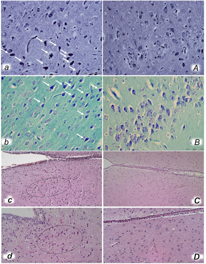
Bielschowsky and Klüver–Barrera histochemical staining presenting neuropathological changes of cerebral cortex in rats with the increased intra-abdominal pressure at 30 mmHg for 30 min (a, A, b, B) treated at 10 min increased intraabdominal pressure time with saline (control a, b) or BPC 157 (A, B). In control rats, an increased number of karyopyknotic cells was found in the cerebral cortex (white arrows) (A, B) that was significantly different from the cortex area in BPC 157-treated rats (a, b). (Bielschowsky staining (a, A); Klüver–Barrera staining (b, B); magnification ×600, scale bar 50 μm). Neuropathological changes of hypothalamic/thalamic area (c, C, d, D) presentation in rats with the increased intra-abdominal pressure at 25 mmHg for 60 min (c, C) or at 50 mmHg for 25 min (d, D), treated at 10 min increased intra-abdominal pressure time with saline (control, c, d) or BPC 157 (C, D). A marked karyopyknosis was found in all control rats (marked in oval) (c, 25 mmHg/60 min); d, 50 mmHg/25 min) while preserved brain tissue was found in BPC 157-treated rats (C, 25 mmHg/60 min); D, 50 mmHg/25 min). (HE; magnification ×400, scale bar 50 μm).

