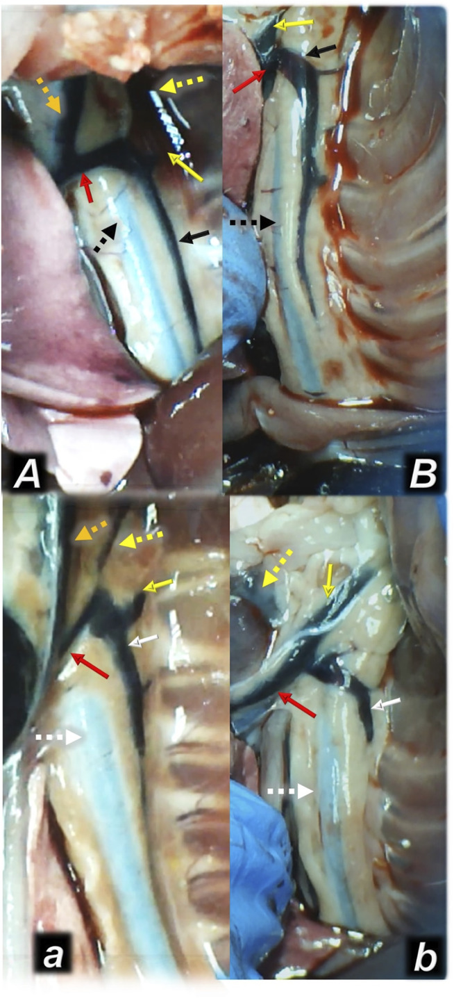
FIGURE 4.

Illustrative presentation of the azygos veins after the increased intraabdominal pressure and medication (sc) (full white arrow, saline (5 ml/kg, low, poor azygos vein presentation a, b) or BPC 157 (full black arrow, 10 ng/kg, upper, functioning azygos vein A, B): 40 mmHg (30 min) (a, A) and 50 mmHg (25 min) (b, B). Aorta (dashed arrows (white (control), black (BPC 157), axillar vein (full yellow arrow), left superior caval vein (red arrow), eternal jugular vein (dashed yellow arrow), internal jugular vein (dark yellow dashed arrow). A camera attached to a VMS-004 Discovery Deluxe USB microscope (Veho, United States).
