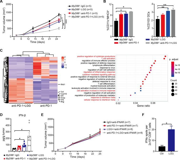
Figure 5.
Lactobacillus rhamnosus GG (LGG) induces a type I interferon (IFN) response via a non-MyD88 dependent manner in dendriticcells (DCs). (A)Myd88 −/− mice were subcutaneously inoculated with MC38 cells and treated with IgG control antibody, anti-PD-1 antibody, oral administration of live LGG (2×10⁹ CFU) or combination of LGG and anti-programmedcell death 1 (PD-1) antibody (n=5). (B) Representative flow cytometry analysis and quantification of DCs and CD103+ DCs in MC38 tumours from Myd88 −/− mice on day 21 given the indicated treatment as described in (A) (n=3). (C) Heatmap illustrating the relative expression of genes and the partitioning of the clusters of genes (gene ontology-GO analysis) based on the RNA-seq analysis of CD11c+ cells in mesenteric lymph nodes (MLN) of MC38 tumour-bearing Myd88 −/− mice given anti-PD-1 antibody or combination treatment with anti-PD-1 antibody and LGG. (D) MC38 tumours in Myd88 −/− mice given the indicated treatment were removed on day 21. The level of IFN-β in tumours was measured using a LEGENDplex cytokine kit. (E)Myd88 −/− mice were subcutaneously inoculated with MC38 cells and treated with IgG control antibody, anti-PD-1 antibody, oral administration of live LGG (2×10⁹ CFU) or a combination of LGG and anti-PD-1 antibody (n=5). Two hundred micrograms anti-IFNAR1 antibody was administered intraperitoneally (i.p.) 24 hours before other treatments. (A and E) Data are expressed as mean±SEM, tumour volume results were analysed by using two-way analysisof variance; *p<0.05; **p<0.01; ***p<0.001. (F) IFN-γ levels in CD8+ T cells from Myd88 −/− mice. The Myd88 −/− mice were gavaged with LGG two times for 1 week. CD11c+ cells were purified from the MLN in and then cocultured with CD8+ T cells and the cross-priming activity of DCs was analysed by IFN-γ ELISPOT assay. (B, D and F) Data are means±SEM; unpaired two-tailed Student’s t-tests; *p<0.05; **p<0.01.

