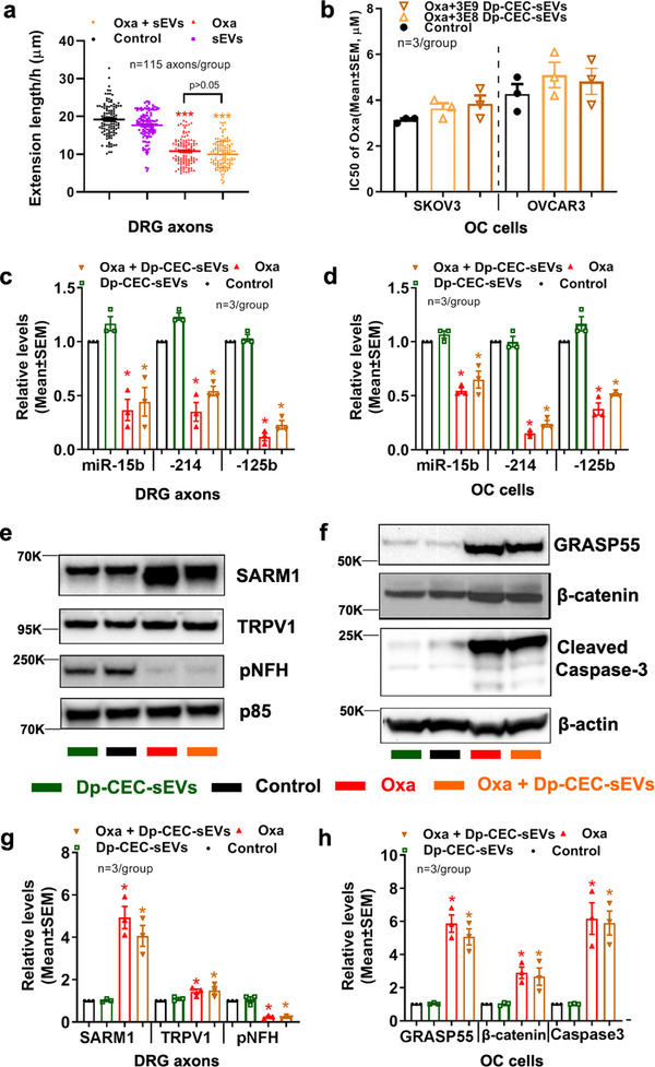
FIGURE 10.

Dp‐CEC‐sEVs do not promote axonal growth and sensitize OC cells to oxaliplatin, and do not alter oxaliplatin‐induced the changes of miRNAs and proteins in axons and OC cells. Quantitative data in a show growth cone extension of DRG axons within 60 minutes and treated with PBS (con), Oxaliplatin (oxa), Dp‐CEC‐sEVs and oxaliplatin in combination with Dp‐CEC‐sEVs (oxa+ Dp‐CEC‐sEVs). Quantitative data in b show IC50 of oxaliplatin in combination with different concentration of Dp‐CEC‐sEVs on SKOV3 and OVCAR3 cells, respectively. qRT‐PCR results show the levels of miR‐15b, 214 and 125b in axons of DRG neurons (c) and in SKOV3 cells (d), respectively, which were received different treatments. Representative Western blots results and their quantitative data show the levels of proteins in axons of DRGs (e, g) and in SKOV3 cells (f, h), respectively, which received different treatments. N in b‐d, g and h indicate the number of replications. K indicates the molecular weight KDa. One‐way ANOVA with Tukey's multiple comparisons test was used. * p<0.05, *** p<0.001 vs control; #, p<0.05 vs oxa. Error bars indicate the standard error of the mean (SEM).
