
FIGURE 1.
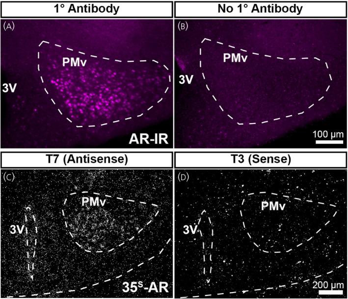
Validation of androgen receptor (AR) immunohistochemistry and Ar in situ hybridization probe. (A, B) Fluorescent images showing AR‐immunoreactivity (AR‐IR) in the adult female mouse brain (postnatal day [PND] 56–70). AR‐IR was observed in sections incubated in primary antibody (A), but not in sections without primary antibody (B). (C, D) Darkfield images showing silver grain deposition corresponding to Ar hybridization signal in adjacent sections from the same brain (PND 12 male mouse). A signal was observed in sections hybridized with an antisense probe (C), but not with a sense probe (D). PMv, ventral premammillary nucleus; 3V, third ventricle. Scale bar = 100 µm (A, B), 200 µm (C, D)