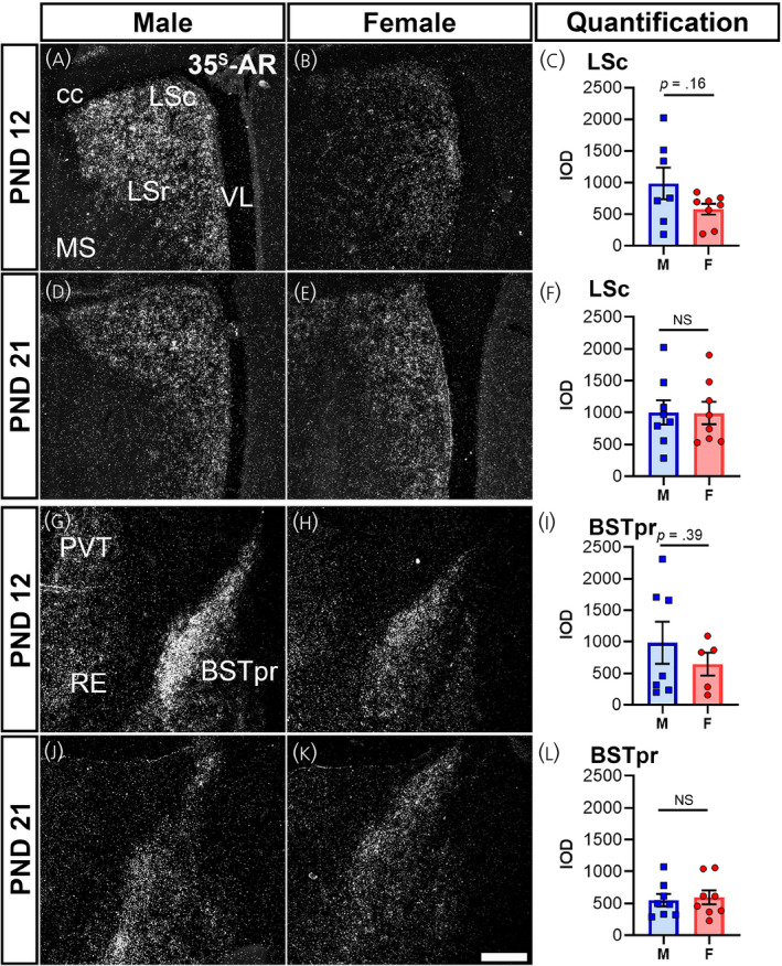
FIGURE 5.

Ar mRNA expression in cerebral nuclei of male and female prepubertal mice. Silver grain deposition corresponding to Ar mRNA hybridization signal in prepubertal postnatal day (PND) 12 (A, B, G, H) and PND 21 (D, E, J, K) male (M) (A, D, G, J) and female (F) (B, E, H, K) mice. (A–F) Lateral septal nucleus, caudodorsal (LSc) and (G–L) bed nucleus of the stria terminalis, principal nucleus (BSTpr). Bar graphs showing the mean ± SEM integrated optical density (IOD) of silver grains (C, F, I, L). IOD was analyzed by a t test with Welch's correction for LSc male vs. female PND 12 (p = .16, n = 7–8 per sex), PND 21 (p = .96, n = 8 per sex), BST male vs. female PND 12 (p = .39, n = 5–7 per sex), and BST male vs. female PND 21 (p = .75, n = 8 per sex). Abbreviations: cc, corpus callosum; LSr, lateral septal nucleus, rostral (rostroventral); MS, medial septal nucleus; PVT, paraventricular nucleus of the thalamus; RE, nucleus of reuniens; VL, lateral ventricle. NS, not significant. Scale bar = 200 µm
