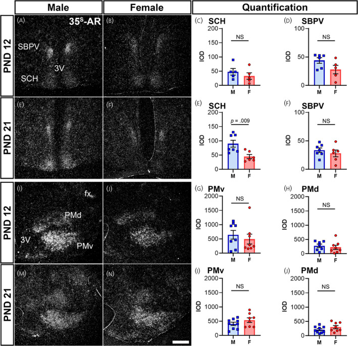
FIGURE 7.

Ar mRNA expression in hypothalamic nuclei of male and female prepubertal mice. Silver grain deposition corresponding to Ar mRNA hybridization signal in prepubertal postnatal day (PND) 12 (A, B, I, J) and PND 21 (E, F, M, N) male (M) (A, E, I, M) and female (F) (B, F, J, N) mice. (A–H) Suprachiasmatic nucleus (SCH) and subparaventricular zone (SBPV), and (I–P) dorsal and ventral premammillary nuclei (PMd and PMv). Note the higher expression of Ar in the SCH of males at PND 21 (E). Bar graphs showing the mean ± SEM integrated optical density (IOD) of silver grains (C, D, G, H, K, L, O, P). IOD was analyzed by a t test with Welch's correction for SCH male vs. female PND 12 (p = .38, n = 5 per sex), SCH male vs. female PND 21 (p = .009, n = 6–7 per sex), SBPV male vs. female PND 21 (p = .45, n = 6–7 per sex), PMv male vs. female PND 21 (p = .21, n = 8–9 per sex), and PMd male vs. female PND 12 (p = .58, n = 7–8 per sex) and PND 21 (p = .19, n = 8–9 per sex), and a Mann–Whitney non‐parametric test for SBPV male vs. female PND 12 (p = .12, n = 6 per sex), and PMv male vs. female PND 12 (p = .57, n = 8 per sex). fx, fornix; 3V, third ventricle. NS, not significant. Scale bar = 200 µm
