
FIGURE 9.
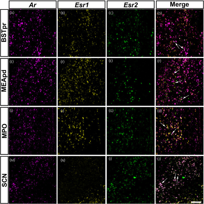
Ar mRNA expression overlaps with Esr1 and Esr2 in specific forebrain nuclei of prepubertal mice. (A–P) Images showing fluorescent in situ hybridization signal for Ar (magenta, A, E, I, M), Esr1 (yellow, B, F, J, N) and Esr2 (green, C, G, K, O). Merge of all three channels shown in (D), (H), (L) and (P). Areas with Ar and Esr1 and/or Esr2 co‐expression include the bed nucleus of the stria terminalis, principal nucleus (BSTpr) (A–D), medial amygdalar nucleus, posterodorsal (MEApd) (E–H), medial preoptic area (MPO) (I–L) and suprachiasmatic nucleus (SCH) (M–P). Arrows show dual or triple‐labeled neurons. Images shown are from postnatal day (PND) 12 female (BSTpr, MEApd, MPO) and male (SCH) mice. Scale bar = 100 µm