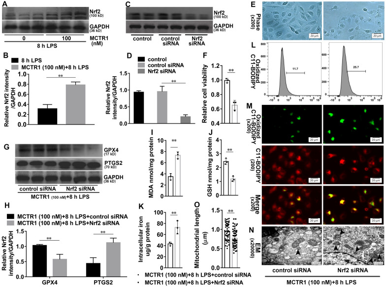
Fig. 5.
Nrf2 is involved in the inhibitory effects of MCTR1 on LPS-induced ferroptosis. A, B HK-2 cells were treated with MCTR1 (100 nM) and LPS (1 ug/ml) for 8 h. Shown are representative western blotting and quantification of Nrf2. HK-2 cells were transfected with 30 nM Nrf2 siRNA for 48 h and then treated with MCTR1 (100 nM) and LPS (1 ug/ml) for 8 h. C, D Representative western blotting and quantification of Nrf2. E Visualization of cell viability were evaluated by phase-contrast microscopy. F Fold change of cell viability. G, H Representative western blotting and quantification of GPX4 and PTGS2. I–K Quantitative analyses of MDA, GSH, and non-heme iron. L Quantitative analyses of oxidized C11-BODIPY 581/591 probe by flow cytometry. M Representative images of C11-BODIPY 581/591 fluorescent ratio-probe. N Representative TEM images. The black arrow indicates ferroptosis-like mitochondria. O Quantitative analyses of mitochondrial length. n = 3, mean ± SD were presented. **P < 0.01

