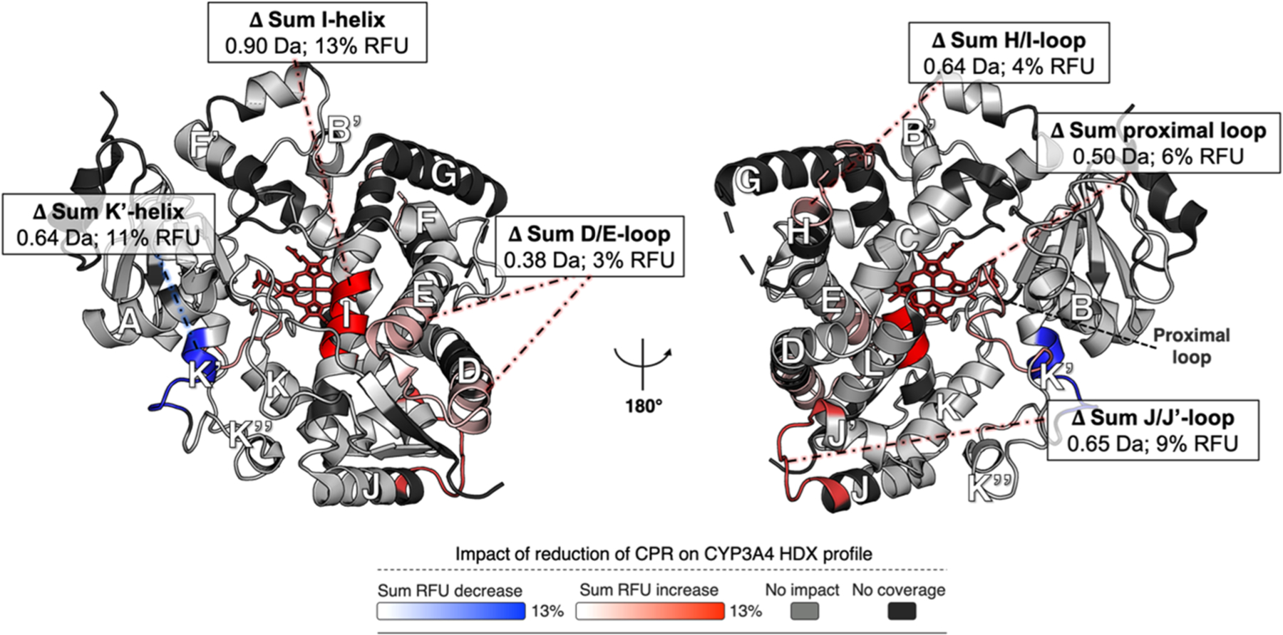
Figure 4.

Impact of the binding of redCPR compared to that of oxCPR on the HDX profile of CYP3A4. The RFU differences were calculated as the uptake of the [CYP:oxCPR] state minus the uptake of the [CYP:redCPR] state. The sum RFU difference of three time points (0.5, 5, and 30 min) is mapped onto the CYP3A4 structure (PDB entry 1W0F), which is shown in both distal (left) and proximal (right) views relative to the heme group (red wireframe). Blue denotes regions that undergo less deuterium uptake upon interaction with redCPR than with oxCPR, whereas red designates regions that undergo more deuterium uptake. The light gray color shows regions for which there is no difference between oxCPR and redCPR binding, and the black regions were not covered by the MS analysis. Overall, the CYP3A4 structure is more dynamic in the presence of redCPR. The sum of deuterium uptake and RFU difference is shown for all statistically significant changes (±0.34 Da, CI of 99%).
