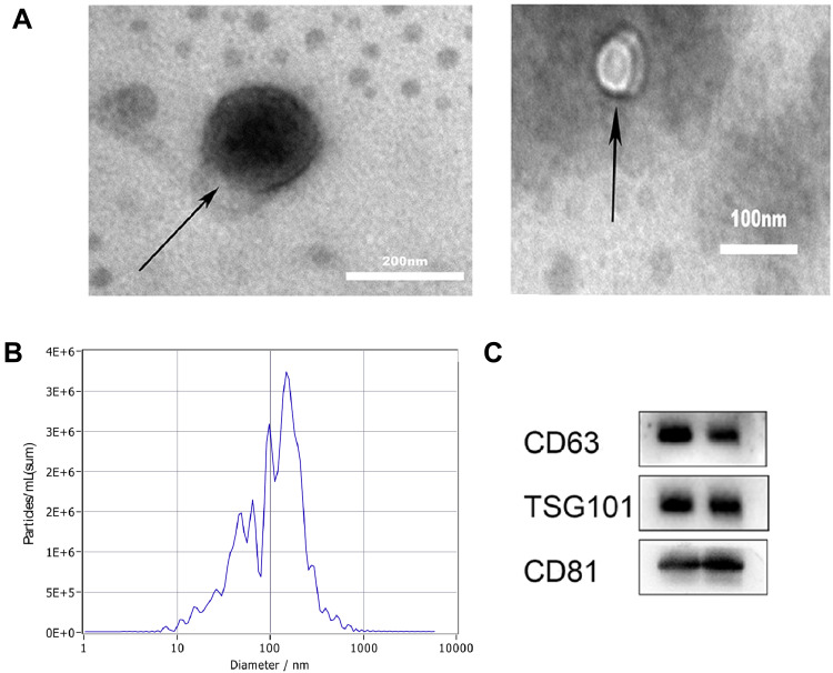
Figure 1.
Characterizing exosomes. (A) Transmission electron microscopy (TEM) images showing the morphology of isolated exosomes. (B) Nanoparticle tracking analysis (NTA) revealed the size distribution and particle concentration of isolated exosomes. (C) Western blot analysis confirmed EV-specific protein marker expression.

