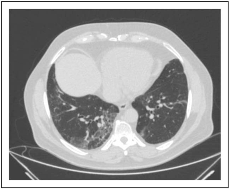
FIGURE 5.
Reticular thickening, bronchiectasis, and ground-gloss opacity. CT of the chest showing inter-intralobular reticular thickening in the right lobe with concomitant thickened wall bronchiectasis bilaterally. Right lower lobe shows GGO-type parenchyma opacity in submantellar location. CT, computed tomography; GGO, ground-gloss opacity.

