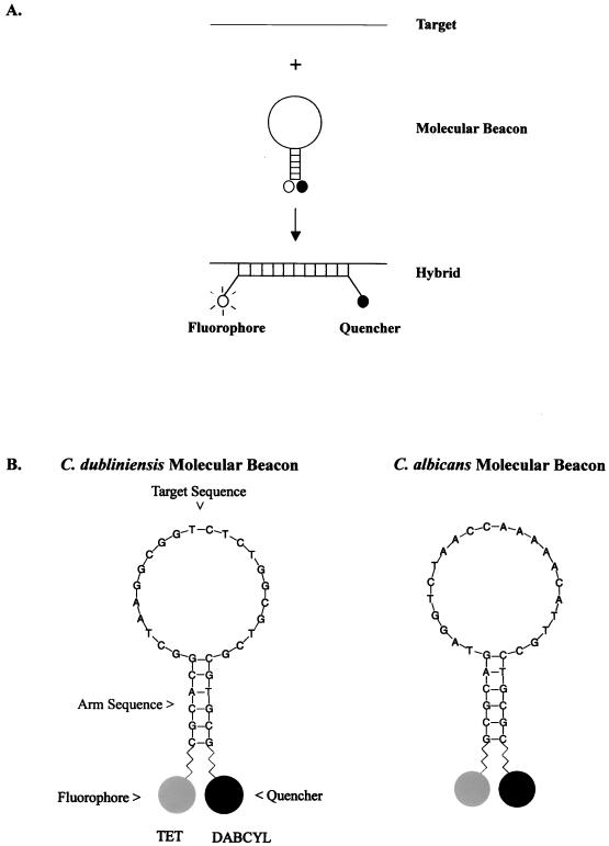
FIG. 1.
(A) Molecular beacon consists of a stem-loop structure with a fluorophore and a quencher bound to the ends of the probe. In free solution, these probes are nonfluorescent because the stem hybrid keeps the fluorophore close to the quencher. When the probe sequence in the loop hybridizes to its target, forming a rigid double helix, a conformational reorganization occurs that separates the quencher from the fluorophore, restoring fluorescence. The figure is adapted from Tyagi and Kramer (51). (B) Nucleotide sequence of the Candida species-specific molecular beacons. The 22-nucleotide target sequence is complementary to the ITS2 region of each Candida species. The fluorophore tetrachloro-6-fluorescein (TET) was attached to the sulfhydryl group on the 5′ arm sequence and 4-(4′-dimethylaminophenylazo)benzoic acid (DABCYL), a quencher, was attached to an amino group on the 3′ arm sequence to form the stem region of the molecular beacon.

