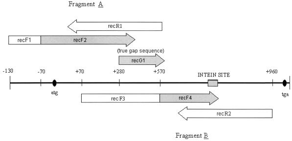
FIG. 1.
Schematic illustration of the primer pairs and sites used in the amplification of the recA gene. The shaded segments correspond to primers applied for seminested PCR. Primer recG1 was used to determine the true sequence of the gap left between primers recR1 and recF4 when seminested PCR was performed.

