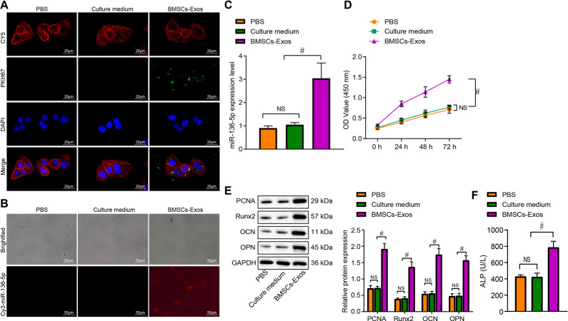
Fig. 3.
miR-136-5p-containing bone marrow mesenchymal stem cell (BMSC)-Exos translocate to MC3T3-E1 and promote its proliferation and osteogenic differentiation. a) BMSC-Exos were labelled with PKH67, and the uptake of BMSC-Exos by MC3T3-E1 was observed under a fluorescence microscope (× 500). The Diluent C labelled cell membrane exhibit red fluorescence, the nuclei exhibit blue fluorescence, and PKH67 labelled Exos exhibit green fluorescence; b) fluorescence microscope observation of the content of Cy3 labelled miR-136-5p in BMSC-Exos in each treatment group with Cy3 exhibiting red fluorescence (× 200); c) reverse transcription quantitative polymerase chain reaction (RT-qPCR) detection of the expression of miR-136-5p in MC3T3-E1 cells treated with BMSC-Exos; d) cell counting kit (CCK)-8 detection of the proliferation rate of MC3T3-E1 cells treated with BMSC-Exos; e) Western blot detection of the protein expression of proliferating cell nuclear antigen (PCNA), Runt-related transcription factor 2 (Runx2), osteocalcin (OCN), osteopontin (OPN), and glyceraldehyde-3-phosphate dehydrogenase (GAPDH) in MC3T3-E1 cells treated with BMSC-Exos; f) alkaline phosphatase (ALP) activity in supernatant of MC3T3-E1 cells treated with BMSC-Exos detected by disodium phosphate microplate method; # indicates p < 0.05, and non-significant (NS) represents p > 0.05. Measurement data were presented as mean and standard deviation. Unpaired t-test was used for data comparison between two groups. Data comparisons among multiple groups were performed using one-way analysis of variance (ANOVA) with Tukey’s post hoc test. Data comparison among groups at different timepoints was performed by repeated measures ANOVA with Bonferroni’s post hoc test. The experiment was repeated three times. NS, non-significant; PBS, phosphate-buffered saline.

