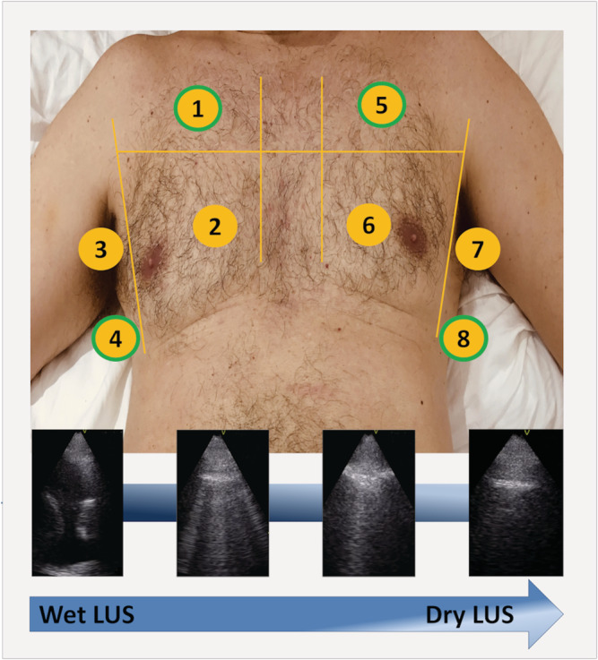
Figure 1.

The eight thoracic areas scanned in the wet‐to‐dry HF study (upper panel): lung areas 1, 4, 5, and 8 (green border) were used for the simplified 4‐lung‐zones protocol; examples of LUS scans showing decreasing lung congestion (lower panel). LUS, lung ultrasound.
