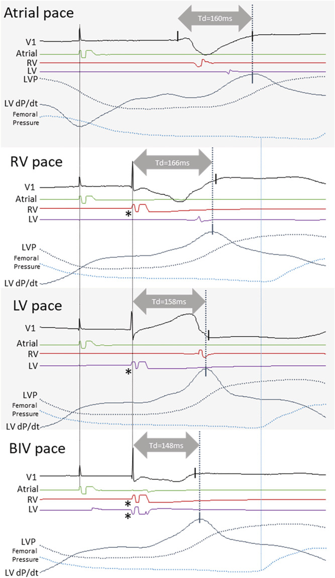
Figure 4.

Synergy from biventricular resynchronization on the timing of peak dP/dt. The panel shows atrial, RV, LV and biventricular pacing with pacing electrode EGMs, LV pressure, LV pressure derivative, and femoral artery pressure from one patient. Td is longer with RV pace than with LV pace, and without myocardial synergy between RV pace and LV pace, one would expect Td with BIV pace (which includes the two pacing electrodes) to be similar to the shorter of that from RV or LV pace; however, BIV pace shortens by 10 ms compared with LV pace demonstrating the presence of myocardial synergy resulting in an earlier pressure increase with BIV pace. Note that a long Q‐LV, RV pace to LV sensed interval, and LV pace to RV sensed interval, confirming the placement of the LV electrode in a late activated region of the LV electrically distant from the RV electrode. Asterisk denotes a pacing artefact. V1, ECG lead; RV, right ventricle; LV, left ventricle; LVP, left ventricular pressure; LV dP/dt, LV pressure derivative; Td, time‐to‐peak LV dP/dt.
