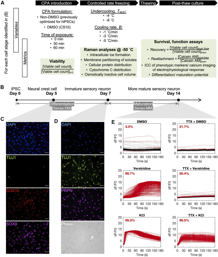
FIGURE 1.
Overview of the cryobiological investigation along a human induced pluripotent stem cell (hiPSC)-to-sensory neuron trajectory. (A) Outline of variables and metrics for the analysis, per neuronal cell stage, of cryoprotective agent (CPA) cytotoxicity before freezing, cell response to variation in nucleation temperature (T NUC ) and cooling ratio (B) during freezing, and cell survival and function after thawing. (B) A 14-day differentiation and maturation timeline for cultivating the three developmental stages of neuronal cells of interest for this investigation. (C) Immunocytochemistry of day-5 neural crest cells (D5 crest) replated at 100,000 cells/cm2, 98.4% TUJ1− SOX9 + SOX10 +. Scale bar: 50 µm. (D) Phase contrast and immunocytochemistry of day-7 sensory neurons (D7 SN) replated at 50,000 cells/cm2, 98.8% TUJ1 + PRPN + excluding dead cells by morphology (example indicated by white arrow in phase). Scale bar: 50 µm. (E) Calcium imaging of day-14 sensory neurons (D14 mSN), seeded at 50,000 cells/cm2 on day 7, showing minimal response to dimethyl sulfoxide (DMSO), strong and tetrodotoxin (TTX)-sensitive response to veratridine, and strong response to potassium chloride (KCl). Lines are color-coded red for responder cells and black for nonresponder cells. Proportion of responder cell population indicated per graph.

