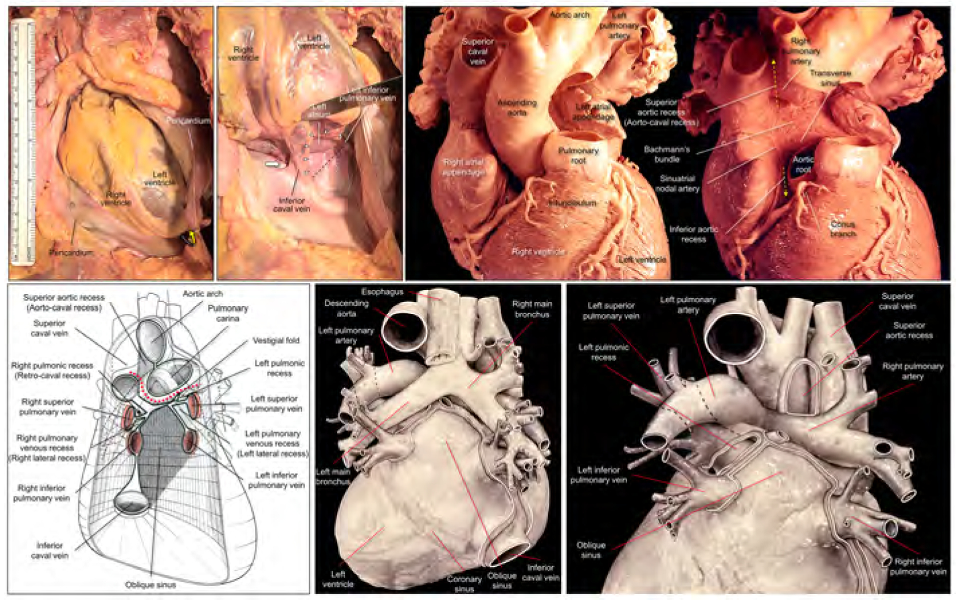
Figure 1. Dissected anatomy of the pericardial space.

Left upper panels show dissection images to show the entrance to the oblique sinus. The apex is raised up (yellow curved arrow) and inverted in the right panel. The entrance of the oblique sinus (white arrowheads and black dashed line), demarcated by the inferior caval vein and the left inferior pulmonary vein is oblique to the vertical body axis. Because of the reflection between the inferior caval vein and the right inferior pulmonary vein, it is impossible to pass this region toward the left side to the oblique sinus (thick white arrow). Right upper panels show high-resolution photographs focusing on the structural anatomy surrounding the transverse sinus recorded by Dr. W.A. McAlpine.18 The pulmonary trunk has been removed in the left panel. Progressive dissection to remove the ascending aorta (right panel) clarifies the space corresponding to the transverse sinus viewed from the slight cranial direction. Refer to Figures 2 and 3. Lower panels are illustrations modified from the McAlpine collection,18 focusing on the pericardial anatomy. Note the oblique entrance of the oblique sinus visualized in the left and middle lower panels. Red dotted line indicates the transverse sinus. Note the hyparterial long left bronchus and eparterial short right bronchus in the middle lower panel (65). Posterior hilum at the roof of the left atrium and its relationship to the extra-pericardial right pulmonary artery can be appreciated in the right lower panel. Illustration courtesy UCLA Cardiac Arrhythmia Center, Wallace A. McAlpine MD collection, reproduced with permission.
